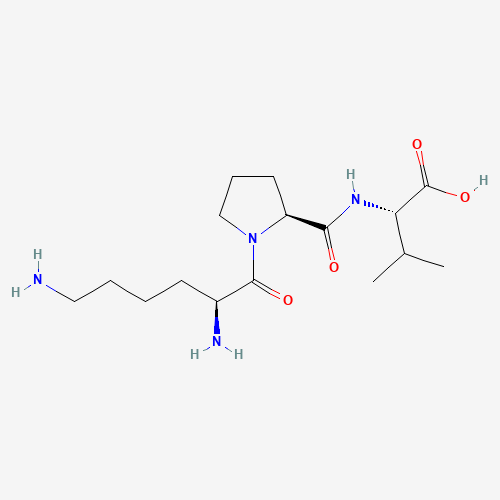
Image 1: Chemical structure of KPV Peptide
In 1989, a study(2) explored the isolation and biological potential of the KPV tripeptide. Upon identifying the COOH terminal peptide in the α-MSH hormone as the primary amino acid messenger sequence, scientists conducted preliminary research to determine if KPV might prevent excessive vasopermeability and swelling of blood vessels. Researchers isolated the KPV peptide and presented it to the experimental mice to assess its potential to mitigate ear swelling. The study concluded that the isolated fragment appeared to inhibit swelling. KPV’s potential anti-inflammatory action may be induced by inactivating inflammatory pathways and possibly inhibiting the synthesis and release of pro-inflammatory cytokines in intestinal and immune cells.
Research
KPV Peptide and Effects on Intestine
As part of a research study,(3) inflamed cells were isolated and exposed to either the KPV peptide or a control. Examination of the cells indicated that even nanomolar concentrations of the peptide appeared to produce anti-inflammatory effects. Researchers suggested that the peptide’s action might be mediated through PepT1 expression in these intestinal cells, implicating PepT1 in transporting the peptide to inflammation sites.
Another study(4) investigated KPV’s potential in addressing ulcerative inflammation of colonic mucosa cells. Researchers hypothesized that KPV might mitigate inflammatory responses within colonic cells by promoting mucosal healing and reducing inflammation. The proposed mechanism involved targeted delivery of KPV to inflamed colonic tissues, where it might exert anti-inflammatory influence. The findings suggested that KPV may protect mucosal surfaces and downregulate TNF-α, a key inflammatory marker.
Further trials(5) using two murine models of intestinal inflammation reported significant improvements following KPV exposure, including earlier recovery, significant weight regain, and reduced inflammatory infiltrates in colonic tissue. These outcomes were supported by a notable decrease in myeloperoxidase (MPO) activity, indicating reduced neutrophil accumulation and inflammation. Additionally, the study explored whether KPV’s anti-inflammatory actions might be linked to the melanocortin-1 receptor (MC1R), leading the researchers to suggest that these effects may be “at least partially independent of MC1R signaling.”
A 1984 study(6) aimed to evaluate the potential antipyretic action of KPV. Rabbits were given the peptide to examine its effects on the nervous system. The results suggested that KPV exhibited antipyretic potential, reducing the rabbits’ body temperature to optimal levels.
KPV Peptide and Intestinal Protection
A study(7) conducted on murine models investigated the potential of KPV peptide in mitigating intestinal inflammation. In this experiment, mice with induced bowel dysfunction were divided into two groups: one receiving the peptide and the other receiving a control. The results indicated that mice exposed to the peptide exhibited a reduction in inflammatory cells and anti-enzymatic symptoms.
Another study on a murine model of intestinal inflammation involved exposure to a chemical-induced compound of KPV and hyaluronic acid. This compound aimed to facilitate the targeted delivery of KPV to specific intestinal locations. The findings indicated a reduction in intestinal swelling, suggesting the peptide’s potential influence in reducing inflammation.
KPV Peptide and Anti-Inflammatory Characteristics
A comparative study(8) was conducted to evaluate the effects of α-MSH and KPV peptides on organ inflammation. In this experiment, mice with induced ear swelling due to skin rashes and dermatitis were divided into two groups. One group received an irritant to induce ear swelling followed by exposure to KPV peptide, while the other group received the irritant followed by α-MSH. After 24 hours, both groups showed similar improvement in reducing ear swelling. After two weeks, researchers ceased exposure to both compounds and continued presenting only the irritant.
The results indicate that the mice exposed to α-MSH continued to exhibit reduced swelling. Researchers noted that, “most of the anti‐inflammatory activities of α‐MSH can be attributed to its C‐terminal tripeptide KPV.”
Mechanisms of Wound Healing and Potential Role of KPV Peptide
Wound healing is a complex biological process involving three main phases: inflammation, proliferation, and remodeling of skin, tissues, or cells. This process is characterized by the presence of various cell types and concentrations of cytokines at the site of injury. Most cells involved in wound healing express the melanocortin 1 receptor (MC1R), where the α-MSH hormone binds. Researchers suggest that analogs of α-MSH, such as the KPV peptide, may also interact with these receptors.(8)
In another study(9) focusing on corneal epithelial wound healing, the potential of KPV peptide was investigated, particularly in relation to nitric oxide (NO) involvement. Following mechanical abrasion to induce damage to the corneal epithelium, varying concentrations of KPV peptide were applied to the tissue. The progress of epithelial wound healing was meticulously monitored and analyzed using computerized software, comparing the mean area of epithelial defects among experimental groups at multiple time points.
The findings indicated a potential acceleration in the healing process of corneal tissues exposed to KPV peptide compared to controls. Specifically, within 60 hours, all corneas exposed to KPV appeared to exhibit complete re-epithelialization, contrasting with the control group where none achieved full healing within the same timeframe.
Further experiments explored the influence of nitric oxide using the inhibitor Nω-nitro-l-arginine methyl ester (l-NAME), which appeared to impede the accelerated healing effect of KPV on corneal epithelial wounds. This suggests that the positive impact of KPV on corneal epithelial wound healing may be linked to nitric oxide dynamics within the tissue.
In vitro experiments with corneal epithelial cells (RCE) exposed to different concentrations of KPV indicated increased cell viability, particularly notable at concentrations of 1 and 10 μM. These results suggest that KPV peptide might not only accelerate corneal epithelial wound healing but also stimulate cell viability, indicating a potential broader reparative role involving nitric oxide modulation.
Scar Recovery
A study was conducted to investigate the potential of KPV peptide in scar recovery using murine models.(10) Juvenile mice were divided into two groups: one group was exposed to KPV, while the other served as a control group. Thirty minutes after exposure, all mice underwent two surgical incisions of 6.5 mm width on their dorsal skin under anesthesia.
Wound healing and scar formation were assessed on days 3, 7, 40, and 60 post-incision. Observations on days 3 and 7 indicated improved skin healing in the peptide-exposed mice, potentially attributed to reduced levels of inflammatory cells such as leukocytes and mast cells. By days 40 and 60, the peptide-exposed mice exhibited smaller scar areas compared to the control group.
KPV Peptide and Tumorigenesis
Studies suggest that KPV peptide may exert inhibitory effects on tumorigenesis in a mouse model of colitis-related cancer. To explore whether KPV might inhibit carcinogenesis independently of inflammation, researchers investigated its impact in APCMin/+ mice, a genetic model prone to intestinal adenocarcinoma. Mice were given KPV for 13 weeks, during which adenomas primarily developed in the small intestine. The findings indicated that KPV did not alter the incidence of tumors in either the small intestine or the colon. However, the data suggested anti-inflammatory characteristics of KPV, as indicated by reductions in intestinal inflammation markers such as lipocalin-2. Despite this apparent anti-inflammatory effect, KPV-induced reduction in inflammation did not appear to mitigate tumor growth in this genetic model of carcinogenesis.(11)
While hereditary colon carcinoma models represent a minority of cases, they exhibit rapid tumor progression in germ-free conditions and significant baseline intestinal inflammation. Thus, although KPV exhibited some potential in attenuating inflammation across various models of colon cancer, its efficacy in suppressing carcinogenesis was limited in the stringent APCMin/+ model. Researchers concluded that “despite modest inhibition of tumorigenesis in such a rigorous model, KPV effectively mitigated inflammation in this model of inflammation-induced carcinogenesis.”
Stomach Cancer
Investigations suggest that KPV, an anti-inflammatory tripeptide transported by PepT1, may offer protective influence against stomach cancer in wild-type mice.
Researchers utilized an AOM/DSS-induced mouse model of colon cancer to assess KPV’s potential inhibitory effects on carcinogenesis. The study(11) suggested that KPV significantly reduced tumor incidence and proliferation of malignant colonic epithelial cells in a PepT1-dependent manner.
NOTE: These products are intended for laboratory research use only. This peptide is not intended for personal use. Please review and adhere to our Terms and Conditions before ordering.
References:
- National Center for Biotechnology Information (2024). PubChem Compound Summary for CID 125672, L-Valine, N-(1-L-lysyl-L-prolyl).
- Hiltz ME, Lipton JM. Antiinflammatory activity of a COOH-terminal fragment of the neuropeptide alpha-MSH. FASEB J. 1989 Sep;3(11):2282-4. https://pubmed.ncbi.nlm.nih.gov/2550304/
- Dalmasso G, Charrier-Hisamuddin L, Nguyen HT, Yan Y, Sitaraman S, Merlin D. PepT1-mediated tripeptide KPV uptake reduces intestinal inflammation. Gastroenterology. 2008 Jan;134(1):166-78. https://pubmed.ncbi.nlm.nih.gov/18061177/
- Xiao, B., Xu, Z., Viennois, E., Zhang, Y., Zhang, Z., Zhang, M., Han, M. K., Kang, Y., & Merlin, D. (2017). Orally Targeted Delivery of Tripeptide KPV via Hyaluronic Acid-Functionalized Nanoparticles Efficiently Alleviates Ulcerative Colitis. Molecular therapy : the journal of the American Society of Gene Therapy, 25(7), 1628–1640. https://doi.org/10.1016/j.ymthe.2016.11.020
- Kannengiesser K, Maaser C, Heidemann J, Luegering A, Ross M, Brzoska T, Bohm M, Luger TA, Domschke W, Kucharzik T. Melanocortin-derived tripeptide KPV has anti-inflammatory potential in murine models of inflammatory bowel disease. Inflamm Bowel Dis. 2008 Mar;14(3):324-31. doi: 10.1002/ibd.20334. PMID: 18092346. https://pubmed.ncbi.nlm.nih.gov/18092346/
- B. Richards, J.M. Lipton, Effect of α-MSH 11–13 (lysine-proline-valine) on fever in the rabbit, Peptides, Volume 5, Issue 4, 1984, Pages 815-817, ISSN 0196-9781, https://doi.org/10.1016/0196-9781(84)90027-5
- Klaus Kannengiesser, MD, Christian Maaser, MD, Jan Heidemann, MD, Andreas Luegering, MD, Matthias Ross, MD, Thomas Brzoska, PhD, Markus Bohm, MD, Thomas A. Luger, MD, Wolfram Domschke, MD, Torsten Kucharzik, MD, Melanocortin-derived tripeptide KPV has anti-inflammatory potential in murine models of inflammatory bowel disease, Inflammatory Bowel Diseases, Volume 14, Issue 3, 1 March 2008, Pages 324–331, https://doi.org/10.1002/ibd.20334
- Brzoska T, Luger TA, Maaser C, Abels C, Böhm M. Alpha-melanocyte-stimulating hormone and related tripeptides: biochemistry, antiinflammatory and protective effects in vitro and in vivo, and future perspectives for the treatment of immune-mediated inflammatory diseases. Endocr Rev. 2008 Aug;29(5):581-602. https://pubmed.ncbi.nlm.nih.gov/18612139/
- Bonfiglio V, Camillieri G, Avitabile T, Leggio GM, Drago F. Effects of the COOH-terminal tripeptide alpha-MSH(11-13) on corneal epithelial wound healing: role of nitric oxide. Exp Eye Res. 2006 Dec;83(6):1366-72. doi: 10.1016/j.exer.2006.07.014. Epub 2006 Sep 11. PMID: 16965771. https://pubmed.ncbi.nlm.nih.gov/16965771/
- de Souza KS, Cantaruti TA, Azevedo GM Jr, Galdino DA, Rodrigues CM, Costa RA, Vaz NM, Carvalho CR. Improved cutaneous wound healing after intraperitoneal injection of alpha-melanocyte-stimulating hormone. Exp Dermatol. 2015 Mar;24(3):198-203. https://pubmed.ncbi.nlm.nih.gov/25431356/
- Viennois E, Ingersoll SA, Ayyadurai S, Zhao Y, Wang L, Zhang M, Han MK, Garg P, Xiao B, Merlin D. Critical role of PepT1 in promoting colitis-associated cancer and therapeutic benefits of the anti-inflammatory PepT1-mediated tripeptide KPV in a murine model. Cell Mol Gastroenterol Hepatol. 2016 May;2(3):340-357. doi: 10.1016/j.jcmgh.2016.01.006. PMID: 27458604; PMCID: PMC4957955. https://www.ncbi.nlm.nih.gov/pmc/articles/PMC4957955/
- Image source: National Center for Biotechnology Information (2024). PubChem Compound Summary for CID 125672, L-Valine, N-(1-L-lysyl-L-prolyl)-. Retrieved July 14, 2024