LL-37 is researched in several fields of study, such as:(1)(2)(3)
- Potential to disrupt microbial metabolism and/or microbial membranes
- Modulatory potential on immune cells
- Modulating cell survival, migration, and angiogenesis
Research
LL-37 and Microbial Structures Disruption
LL-37 is posited to negatively interact with microbial organisms by disrupting their membrane structures and forming pores. For example, when interacting with Escherichia coli ML-35p, LL-37 was observed to potentially permeabilize both the inner and outer membranes, suggesting that once it inserts into or traverses the outer membrane, it may rapidly compromise the cytoplasmic membrane as well. According to a study by Turner et al., cationic charges on LL-37 may contribute to this observable interaction with the bacterial surface components. (4} This may be attributable to the electrostatic attraction between LL-37 and the anionic membrane that enables the peptide to nestle into, or even span, bacterial membranes. This process might be facilitated by the peptide’s potential binding to LPS. Indeed, the authors posited that LL-37 displays positive cooperativity (a Hill coefficient near 2) in its binding to LPS. This can be understood to mean that two LL-37 molecules may interact with each LPS molecule in a mutually reinforcing manner.
This cooperative binding presumably helps LL-37 cluster or locally reorganize bacterial surface elements, which may support membrane permeabilization. Although LL-37 was posited to indicate broad-spectrum potential under varied conditions, the researchers also suggested that particular microbes, such as Burkholderia cepacia, may resist the peptide’s actions in the study’s assays, possibly because these research models possess outer membrane modifications or other protective factors that limit the peptide’s access to, or insertion into, the bilayer. The researchers also highlighted the potential for using various cultural media as an important technical consideration.
Certain standard broths, many of which are rich in polyanionic components, led to lower observed activity for LL-37, presumably because those anionic fragments might sequester or precipitate cationic peptides. The researchers indicated that when the broth was refined through anion exchange to reduce these confounding components, LL-37 more readily maintained its bactericidal behavior in vitro. Some studies also suggest that the peptide may exhibit activity against enveloped viruses such as HSV-1 (herpes simplex virus) vaccinia virus and may inhibit non-enveloped viruses (e.g., certain adenovirus strains) through different mechanisms (e.g., disrupting viral envelopes or interfering with viral entry). ( 3)
LL-37 and Immune Cell Regulation
LL-37 is believed to have the potential to recruit a variety of immune cells, such as neutrophils, monocytes, and T cells. For example, research by Sadek et al. suggests that LL-37 may influence neutrophils when they are exposed to microbial stimuli in vitro. (5 ) More specifically, it is posited that LL-37 may increase the production of reactive oxygen species (ROS) in neutrophils, apparently through mechanisms involving the peptide’s ability to support the intracellular pathways tied to the respiratory burst. Respiratory burst is the process by which phagocytic cells consume large amounts of oxygen during phagocytosis. Thus, the LL-37 peptide may facilitate the uptake of mediator molecules that may intensify ROS production. Moreover, the addition of LL-37 was suggested to play some role in augmenting the engulfment of bacteria during phagocytosis.
These authors and others have posited that these actions may contribute to bolstering the microbicidal abilities of these cells. To research the role of LL-37 further, investigating scientists employed neutrophils from murine models a. They observed that these cells may release more TNF-α upon bacterial stimulation while exhibiting lower bactericidal capacity compared to wild-type controls. This suggests that CRAMP—or LL-37 in other contexts—potentially helps to fine-tune the balance between the killing of pathogens and the control of excessive inflammatory signaling. The presence of the precursor and active forms of cathelicidin in supernatants from unstimulated and stimulated neutrophils supports the idea that cathelicidin is constitutively available and may be released in response to microbial cues.
According to Takahashi et al., LL-37 might also activate immune cells by interacting with scavenger receptors on their surface and mediating their interaction with regulatory molecules like self-RNA.(6) Notably, the interaction between LL-37 and scavenger receptors such as SR-A6 and SR-B1 may be essential for this process, as blocking these receptors with a competitive inhibitor like fucoidan or silencing their expression may significantly reduce cytokine production. This potentially enables self-RNA to activate intracellular pathways that lead to inflammatory cytokine production (for instance, interleukin-6 and interferon-β1).
Another experiment by Kusaka et al. suggests that once LL-37 disrupts microbial structures like LPS, it may consequently reduce proinflammatory signaling to limit collateral damage. (7) More specifically, the disruption of the LPS structures by LL-37 has been observed to be followed by a diminished induction of key proinflammatory mediators, including IL-6 and IL-8, suggesting that LL-37 may exert a local immunomodulatory influence in inflamed tissues. Thus, Kusaka et al. also posited that, by interfering with LPS, LL-37 might potentially limit excessive inflammatory responses. Moreover, in the presence of tissue injury or necrosis, the release of nucleic acids might further engage TLR-3, possibly amplifying LL-37’s antimicrobial and immunomodulatory activities.
LL-37 and Blood Vessel Growth
SaSalvadort al. have posited that LL-37 may act as a multifunctional polypeptide that may induce a blood vessel-promoting response in endothelial cells via a pathway involving the biosynthesis and downstream signaling of prostaglandin E2 (PGE2). (8 ) The researchers commented that “ LL-37 triggers PGE2 synthesis in endothelial cells in a concentration-dependent manner with maximal induction after 4 hours.” To achieve this in laboratory settings, LL-37 apparently increases intracellular calcium levels and triggers phosphorylation of cytosolic phospholipase A2 (cPLA2) in endothelial cells, possibly resulting in greater release of arachidonic acid substrates. This process may be linked to the concomitant phosphorylation of mitogen-activated protein kinases, particularly Erk, which is considered a pivotal step in cPLA2 activation. Once active, cPLA2 may release arachidonic acid in sufficient amounts to feed cyclooxygenase activity.
Although both cyclooxygenase (COX)-1 and COX-2 enzymes may be present in endothelial cells, COX-1 is considered to play the primary role in generating LL-37–induced PGE2. Various pharmacological inhibitors and gene silencing approaches apparently indicate that blocking COX-1 function abolishes the PGE2 increase in these cultures, whereas selective inhibition of COX-2 has a more modest action on LL–37–stimulatedGE2 production. This outcome aligns with observations that the cord-like structures formed by endothelial cells on a matrix scaffold, in the presence of LL-37, may be diminished when COX-1 is inhibited. Adding exogenous PGE2 back into the system may potentially rescue the angiogenic phenotype, suggesting that LL-37’s capacity to promote endothelial tubule formation is at least partly due to CCOX–1–derived PGE2.
Additional in vitro experiments suggest that LL–37–induced PGE2 may act predominantly through EP3 receptors to promote angiogenesis-like processes. Selective antagonism of this receptor apparently reduces LL–37–stimulated endothelial cord formation, whereas blocking other EP receptor subtypes leads to less pronounced changes. This observation possibly highlights EP3 signaling as a central conduit for the proangiogenic signals originating from PGE2 in these cells.
LL-37 and Wound Tissue Repair
Thanks to its aforementioned proangiogenic potential, Ramos et al. have also posited that “LL37 may have a key role in wound regeneration through vascularization.”.(9) Notably, in vitro assessments suggest that LL-37 might stimulate proliferation, migration, and the formation of tubule-like structures in endothelial cell lines. Such observations have led researchers to hypothesize that this peptide may contribute to angiogenesis. These actions are possibly mediated by formyl peptide receptor-like 1 (FPRL-1), given that LL-37 may apparently engage this receptor to induce cellular signaling cascades and calcium flux, which may foster new vessel formation. In a dexamethasone-compromised wound healing model, LL-37 apparently better-supported re-epithelialization and promoted the appearance of blood vessels in the wound area.
Dexamethasone is often exposed to research models to impair wound repair, so these results may indicate that the peptide’s immunomodulatory and proangiogenic properties help overcome glucocorticoid-mediated suppression of tissue regeneration. While the exact pathway through which LL-37 exerts this vascular and regenerative action remains to be fully clarified, it is speculated that the peptide’s ability to encourage inflammatory balance, recruit or activate cells involved in repair, and support vascularization might all combine to facilitate more robust tissue restoration in such models.
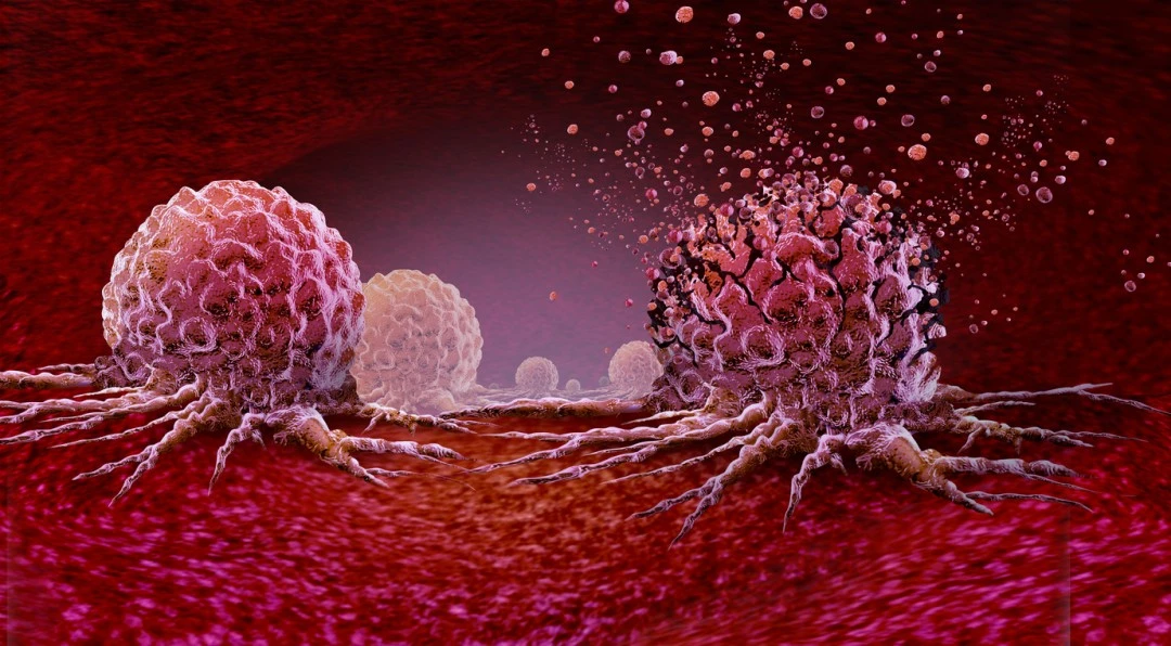
LL-37 and Cancer Cells
Research by Lu et al. suggests that LL-37 may also interact with cancer cells in vitro settings in a variety of ways. (10) Notably, the peptide triggers cell death through a non-membranolytic, receptor-linked mechanism, which may engage G protein-coupled receptors. Some data indicates that LL-37 might modulate intracellular pathways associated with p53, Bcl-2, and Bax/Bak, apparently leading to apoptosis. There are observations that the peptide may upregulate pro-apoptotic effectors (e.g., Bax or p53-upregulated modulator of apoptosis, abbreviated as PUMA) while simultaneously downregulating anti-apoptotic proteins (e.g., Bcl-2). This may culminate in either caspase-dependent or caspase-independent apoptotic signaling, possibly involving the translocation of apoptosis-inducing factor (AIF) or endonuclease G (EndoG).
A further scenario highlighted by Lu et al. suggests that LL-37 might regulate intracellular protein degradation pathways by inhibiting proteasome function, leading to the accumulation of pro-differentiation or tumor-suppressive signals. Researchers have posited that a reduction in proteasome activity might upregulate bone morphogenetic protein 4 (BMP4) and other components of BMP signaling, thus arresting the cell cycle in G0/G1 by modifying levels of p21 and cyclin E2. This may explain why LL-37-exposed cells in certain systems seemed to display less proliferative activity, hinting at a potential interplay between peptide-mediated proteasome inhibition and key cell-cycle checkpoints.
Researchers have also posited that LL-37 might support the activity of immune cells against aberrant or transformed cells. Nevertheless, LL-37’s potential may not be universally inhibitory. Lu et al. commented that other investigators have reported observations where LL-37 apparently promoted proliferation in some tumor cell lines, perhaps via Wnt/β-catenin signaling or by influencing NF-κB phosphorylation. Why LL-37 might have opposing outcomes in different experimental cancer models is still debated. There is not yet any definitive explanation for this.
You can find LL-37 for sale with 99% purity, on our website (available for research use only).
NOTE: These products are intended for laboratory research use only. This peptide is not intended for personal use. Please review and adhere to our Terms and Conditions before ordering.
References:
- Kahlenberg JM, Kaplan MJ. Little peptide, big effects: the role of LL-37 in inflammation and autoimmune disease. J Immunol. 2013 Nov 15;191(10):4895-901. doi: 10.4049/jimmunol.1302005. PMID: 24185823; PMCID: PMC3836506.
- Seil M, Nagant C, Dehaye JP, Vandenbranden M, Lensink MF. Spotlight on Human LL-37, an Immunomodulatory Peptide with Promising Cell-Penetrating Properties. Pharmaceuticals (Basel). 2010 Nov 1;3(11):3435–60. doi: 10.3390/ph3113435. PMCID: PMC4034075.
- Gordon YJ, Huang LC, Romanowski EG, Yates KA, Proske RJ, McDermott AM. Human cathelicidin (LL-37), a multifunctional peptide, is expressed by ocular surface epithelia and has potent antibacterial and antiviral activity. Curr Eye Res. 2005 May;30(5):385-94.doi: 10.1080/02713680590934111. PMID: 16020269; PMCID: PMC1497871.
- Turner J, Cho Y, Dinh NN, Waring AJ, Lehrer RI. Activities of LL-37, a cathelin-associated antimicrobial peptide of human neutrophils. Antimicrob Agents Chemother. 1998 Sep;42(9):2206-14. doi: 10.1128/AAC.42.9.2206. PMID: 9736536; PMCID: PMC105778.
- Alalwani SM, Sierigk J, Herr C, Pinkenburg O, Gallo R, Vogelmeier C, Bals R. The antimicrobial peptide LL-37 modulates the inflammatory and host defense response of human neutrophils. Eur J Immunol. 2010 Apr;40(4):1118-26. doi: 10.1002/eji.200939275. PMID: 20140902; PMCID: PMC2908514.
- Takahashi T, Kulkarni NN, Lee EY, Zhang LJ, Wong GCL, Gallo RL. Cathelicidin promotes inflammation by enabling the binding of self-RNA to cell surface scavenger receptors. Sci Rep. 2018 Mar 5;8(1):4032. doi: 10.1038/s41598-018-22409-3. PMID: 29507358; PMCID: PMC5838106.
- Kusaka S, Nishida A, Takahashi K, Bamba S, Yasui H, Kawahara M, Inatomi O, Sugimoto M, Andoh A. Expression of human cathelicidin peptide LL-37 in inflammatory bowel disease. Clin Exp Immunol. 2018 Jan;191(1):96-106. doi: 10.1111/cei.13047. Epub 2017 Sep 28. PMID: 28872665; PMCID: PMC5721246.
- Salvado MD, Di Gennaro A, Lindbom L, Agerberth B, Haeggström JZ. Cathelicidin LL-37 induces angiogenesis via PGE2-EP3 signaling in endothelial cells, in vivo inhibition by aspirin. Arterioscler Thromb Vasc Biol. 2013 Aug;33(8):1965-72. doi: 10.1161/ATVBAHA.113.301851. Epub 2013 Jun 13. PMID: 23766266.
- Ramos R, Silva JP, Rodrigues AC, Costa R, Guardão L, Schmitt F, Soares R, Vilanova M, Domingues L, Gama M. Wound healing activity of the human antimicrobial peptide LL37. Peptides. 2011 Jul;32(7):1469-76. doi: 10.1016/j.peptides.2011.06.005. Epub 2011 Jun 13. PMID: 21693141.
- Lu F, Zhu Y, Zhang G, Liu Z. Renovation as innovation: Repurposing human antibacterial peptide LL-37 for cancer therapy. Front Pharmacol. 2022 Aug 23;13:944147. doi: 10.3389/fphar.2022.944147. PMID: 36081952; PMCID: PMC9445486.