
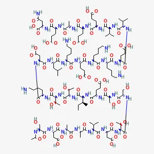
Fig 1 – Thymosin Alpha-1 Chemical Structure
Latest Research on Tα1 Peptide
Thymosin Alpha-1 and Immune Cell Maturation
Dominari et al. proposed that Thymosin Alpha-1 peptide may influence the differentiation and maturation of T cells, particularly CD4+ helper T cells and CD8+ cytotoxic T cells, impacting both innate and adaptive immune responses. This influence likely occurs through Tα1’s interaction with toll-like receptors (TLRs) on antigen-presenting cells. Specifically, the researchers commented that “Thymosin alpha 1 functions as a toll-like receptor (TLR)-9 and TLR-2 agonist in both myeloid and dendritic cells, the professional antigen-presenting cells.” TLRs are a class of proteins that recognize pathogen-associated molecular patterns, triggering immune responses. When Thymosin Alpha-1 peptide binds to TLRs, it may stimulate the production of cytokines—signaling proteins that regulate immune responses—and enhance the antigen-presenting capabilities of these cells. This process is considered to be critical for initiating and shaping immune responses.
For example, by interacting with TLRs on dendritic cells, Thymosin Alpha-1 peptide is thought to enhance their phagocytic ability—allowing them to engulf pathogens—and their capacity for antigen presentation. In murine models, Thymosin Alpha-1 peptide has appeared to have an ability to modulate the activity of natural killer (NK) cells and CD8+ T cells, both of which are crucial for identifying and eliminating infected or tumor cells. This suggests that Tα1 may enhance the immune system’s capacity to target and destroy abnormal cells.(3)
Tao et al. further explored the specific molecular mechanisms by which Thymosin Alpha-1 peptide may potentially influence immune cell maturation. Their research suggests that Tα1 may bind to TLR3, TLR4, and TLR9, which in turn activates intracellular signaling pathways such as IRF3 (interferon regulatory factor 3) and NF-κB (nuclear factor kappa-light-chain-enhancer of activated B cells). These pathways are posited to drive the proliferation and activation of immune cells, influencing their development and function. Other TLRs, such as TLR2 and TLR7, might also play a role in Tα1’s activity, triggering additional pathways like TLR2/NF-κB, TLR2/p38 MAPK (mitogen-activated protein kinase), and TLR7/MyD88 (myeloid differentiation primary response 88). These signaling pathways are integral to cytokine production, further supporting immune cell maturation.(4)
Thymosin Alpha-1 and Immune Cell Differentiation
Based on Tao et al.’s research, one of Tα1’s roles may include promoting the differentiation of precursor T cells into cytotoxic T lymphocytes (CTLs), which are deemed essential for the immune system’s ability to eliminate infected cells. By binding to TLRs on these precursor cells, Thymosin Alpha-1 peptide might support the generation of CD8+ T cells, increasing their potential to recognize and destroy infected cellular targets. Tα1 also appears to be involved in the differentiation of dendritic cells, which are considered key antigen-presenting cells that activate T cells. During this process, Tα1 may increase the expression of surface activation markers, which may be critical for T-cell stimulation. It may also influence the polarization of T-helper cells, potentially favoring the differentiation of certain T-helper cell subsets depending on the cytokine environment.(4)
Thymosin Alpha-1 and Immune Cell Response Regulation
Dominari et al., have also suggested that Tα1 might induce the production of key immune mediators such as interleukins (IL-2, IL-10, IL-12), interferons (IFN-α and IFN-γ), and other cytokines, which are believed to be important for regulating immune responses.(3) The peptide’s potential to stimulate these cytokines has led to hypotheses that it might be beneficial in immunodeficient models, as well as in controlling excessive immune responses, such as cytokine storms. For example, it is posited that Tα1 could have a role in reversing T cell depletion seen in suppressed immune cell functioning. In particular, the authors commented that the peptide may also have beneficial effects in lab models of “suppressed lymphokine-activated killer cell activity and […] immunodeficiency.”(3)
On the other hand, Dominari et al. highlighted that some studies using murine (mouse) models suggest that thymosin alpha-1 (Tα1) may reduce levels of interleukin-1 beta (IL-1β) and TNF-α, two key inflammatory cytokines.(3) These reductions may lead to decreased inflammatory responses, offering potential protection in conditions characterized by excessive inflammation, such as cytokine storm models.
For instance, Matteucci et al. observed that Tα1 may mitigate cytokine dysregulation, which is often seen in hyperinflammatory conditions. In experiments using proinflammatory stimuli like lipopolysaccharides (LPS), Tα1 exposure resulted in CD8+ T cells suggesting reduced production of certain cytokines.(5) These downregulated genes are associated with pathways such as IL-17 and TLR signaling, both of which are critical to initiating and maintaining inflammatory responses. This implies that Tα1 may suppress the pathways typically activated during cytokine storms.
Additionally, Tα1 appears to influence the expression of activation markers such as CD38 and HLA-DR in CD8+ T cells. In murine models simulating inflammatory conditions, Tα1 reduced the expression of these markers, suggesting its potential role in calming overly active immune responses. However, these actions seem to be context-dependent, with Tα1 regulating immune pathways differently depending on whether the environment is inflammatory or not.
Furthermore, Tα1 has been suggested to upregulate interleukin-10 (IL-10), an anti-inflammatory cytokine, which could further contribute to its immunomodulatory actions. This indicates that Tα1 might have a dual role: suppressing proinflammatory cytokines while promoting cytokines that help restore immune balance. Tα1’s modulation of cytokine-related pathways, including nuclear factor kappa B (NF-κB) and p38 mitogen-activated protein kinase (p38 MAPK), suggests it may interact with TLR signaling, a key mechanism that detects infected or damaged cells. This could explain why Thymosin Alpha-1 peptide has been observed to reduce inflammatory responses without compromising the overall immune function.(5)
Thymosin Alpha-1 and Tumor Cells
As summarized by Dominari et al., several studies in murine studies suggest that Thymosin Alpha-1 peptide may reduce tumor growth through either direct action on tumor cells or by modulating the immune environment to better target tumor cells.(3)
Kharazmi-Khorassani et al. suggest that a notable function of Tα1 may be its inhibition of tumor cell migration. Studies using scratch assays indicated that Th1 potentially inhibits the migratory capacity of A549 cells, reducing migration by about 20-26.5% after 48 hours. This anti-migratory action might implicate Thα1 in modulating cellular pathways involved in tumor cell metastasis, although the underlying mechanisms remain speculative.(6)
Additionally, Thymosin Alpha-1 peptide may influence the tumor microenvironment by modulating immune infiltration and enhancing antigen presentation. Studies have suggested that Tα1 may induce the expression of major histocompatibility complex (MHC) class I on tumor cells, potentially increasing their visibility to T cells. This function might help “prime” the immune system for subsequent laboratory interventions against tumor cells, thereby promoting a more robust antitumor response.
In particular, Thymosin Alpha-1 peptide has been suggested to increase immune cell infiltration, including CD4+ and CD8+ T cells, around tumor cells. The immune maintenance role of Thymosin Alpha-1 peptide is another area where its long-term actions on tumor cell suppression have been considered. It has been posited that Tα1 may contribute to sustaining immune surveillance even after the initial response, helping to prevent tumor cell recurrence. This could be due to its influence on immune homeostasis, particularly by modulating the balance between effector T cells and regulatory T cells. Moreover, Tα1 has been suggested to possibly protect against immune-mediated tissue damage which might be relevant in preventing collateral damage during immune cell responses to tumor cells.(7)
Thymosin Alpha-1 and Antioxidative Potential
Additionally, Dominari et al. suggest that Thymosin Alpha-1 may have the ability to reduce oxidative stress by enhancing antioxidant enzyme activities (e.g., superoxide dismutase and glutathione peroxidase). Via this mechanism, Thymosin Alpha-1 peptide might protect various types of somatic cells from damage according to suggestions from different animal models.(3)
Based on research by Kharazmi-Khorassani et al., Thymosin Alpha-1 peptide has been suggested to possess potential antioxidant properties, as detailed by its potential to modulate the activity of critical antioxidant enzymes. In A549 cells, Tα1 has apparently enhanced the activities of catalase, superoxide dismutase (SOD), and glutathione peroxidase (GPx) in a concentration-dependent manner. This enhancement of antioxidant enzyme activities may contribute to Thα1’s ability to reduce reactive oxygen species (ROS) levels, as seen in research models.(6)
Thymosin Alpha-1 and Immune-Nerve Cell Interactions
Research conducted by Wang et al. indicates that Tα1 may have immunomodulatory actions that influence neurodevelopment. In murine models, Thymosin Alpha-1 peptide has been reported by researchers to increase the expression of neural progenitor markers, such as nestin and T-box brain gene 2 (Tbr2), as well as newly formed nerve cells, which are identified by the presence of BrdU and NeuN-positive cells. This action is especially notable in the hippocampus, a brain region considered critical for learning and memory. These observations suggest that Tα1 may play an important role in promoting the early stages of nerve cell formation and maturation.
A possible mechanism underlying Tα1’s role in neurogenesis involves its ability to shift the immune system towards a T-helper 1 (Th1) cell response. This Th1 bias is characterized by increased levels of cytokines such as interferon-gamma (IFN-γ) and interleukin-4 (IL-4), both of which are associated with enhanced neurogenesis and cognitive function. These cytokines may promote the proliferation of neuronal cells, particularly within the hippocampus. In addition, Tα1 appears to reduce the levels of pro-inflammatory cytokines like interleukin-6 (IL-6) and TNF-α, which have been linked to neuroinflammation and cognitive decline. By decreasing inflammation and fostering a more neurotrophic environment, Tα1 may potentially protect neural tissues and support nerve cell function.
Another important aspect of Tα1’s action may involve its regulation of neurotrophic factors—proteins that may support the growth and survival of nerve cells. Studies suggest that Thymosin Alpha-1 peptide increases the expression of brain-derived neurotrophic factor (BDNF), nerve growth factor (NGF), and insulin-like growth factor-1 (IGF-1) in the hippocampus. These neurotrophic factors are considered to be essential for neuronal survival, growth, and synaptic plasticity. Their upregulation in response to Thymosin Alpha-1 peptide may explain the improved cognitive performance and neuroplasticity observed in test animals. The interplay between the IFN-γ/IL-4 ratio and the levels of neurotrophic factors further suggests that the immune modulation triggered by Thymosin Alpha-1 peptide is intricately linked to its neurogenic actions. By modulating both immune responses and neurotrophic signaling, Tα1 appears to create an environment that supports nerve cell development and function.(8)
NOTE: These products are intended for laboratory research use only. This peptide is not intended for personal use. Please review and adhere to our Terms and Conditions before ordering.
References:
- Pierluigi, B., D’Angelo, C., Fallarino, F., Moretti, S., Zelante, T., Bozza, S., De Luca, A., Bistoni, F., Garaci, E., & Romani, L. (2010). Thymosin alpha1: the regulator of regulators?. Annals of the New York Academy of Sciences, 1194, 1–5. https://doi.org/10.1111/j.1749-6632.2010.05465.x
- Li, J., Liu, C. H., & Wang, F. S. (2010). Thymosin alpha 1: biological activities, applications and genetic engineering production. Peptides, 31(11), 2151–2158. https://doi.org/10.1016/j.peptides.2010.07.026
- Dominari, A., Hathaway Iii, D., Pandav, K., Matos, W., Biswas, S., Reddy, G., Thevuthasan, S., Khan, M. A., Mathew, A., Makkar, S. S., Zaidi, M., Fahem, M. M. M., Beas, R., Castaneda, V., Paul, T., Halpern, J., & Baralt, D. (2020). Thymosin alpha 1: A comprehensive review of the literature. World journal of virology, 9(5), 67–78. https://doi.org/10.5501/wjv.v9.i5.67
- Tao, N., Xu, X., Ying, Y., Hu, S., Sun, Q., Lv, G., & Gao, J. (2023). Thymosin α1 and Its Role in Viral Infectious Diseases: The Mechanism and Clinical Application. Molecules (Basel, Switzerland), 28(8), 3539. https://doi.org/10.3390/molecules28083539
- Matteucci, C., Minutolo, A., Balestrieri, E., Petrone, V., Fanelli, M., Malagnino, V., Ianetta, M., Giovinazzo, A., Barreca, F., Di Cesare, S., De Marco, P., Miele, M. T., Toschi, N., Mastino, A., Sinibaldi Vallebona, P., Bernardini, S., Rogliani, P., Sarmati, L., Andreoni, M., Grelli, S., … Garaci, E. (2020). Thymosin Alpha 1 Mitigates Cytokine Storm in Blood Cells From Coronavirus Disease 2019 Patients. Open forum infectious diseases, 8(1), ofaa588. https://doi.org/10.1093/ofid/ofaa588
- Kharazmi-Khorassani, J., & Asoodeh, A. (2019). Thymosin alpha-1; a natural peptide inhibits cellular proliferation, cell migration, the level of reactive oxygen species and promotes the activity of antioxidant enzymes in human lung epithelial adenocarcinoma cell line (A549). Environmental toxicology, 34(8), 941–949. https://doi.org/10.1002/tox.22765
- Costantini, C., Bellet, M. M., Pariano, M., Renga, G., Stincardini, C., Goldstein, A. L., Garaci, E., & Romani, L. (2019). A Reappraisal of Thymosin Alpha1 in Cancer Therapy. Frontiers in oncology, 9, 873. https://doi.org/10.3389/fonc.2019.00873
- Wang, G., He, F., Xu, Y., Zhang, Y., Wang, X., Zhou, C., Huang, Y., & Zou, J. (2017). Immunopotentiator Thymosin Alpha-1 Promotes Neurogenesis and Cognition in the Developing Mouse via a Systemic Th1 Bias. Neuroscience bulletin, 33(6), 675–684. https://doi.org/10.1007/s12264-017-0162-x
- Figure 1: https://pubchem.ncbi.nlm.nih.gov/image/imgsrv.fcgi?cid=16130571&t=l